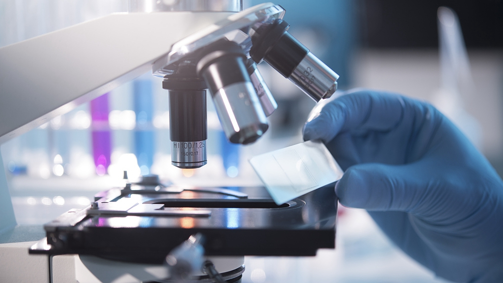
1. Nat Commun: Bioengineering micro-cocoon protects drug molecules with amazing effects!

For scientists, finding inspiration from nature is a common means of human life. In the past, scientists have been inspired by the enzymatic reactions in nature to produce energy, and are inspired by cactus to store water. Today, we use the natural characteristics of silkworm cocoons to create a carrier suitable for small molecules. “Many drugs have excellent therapeutic effects, but they are less stable and difficult to store. This is a common problem in medical practice,” said Tuomas Knowles, lead author of the study. Therefore, they hope that silkworm cocoons can help prolong the life of drug molecules. Because silk is a biodegradable residue and has a low production cost, it has been used to make other surgical supplies, including brain implants, optical devices, Xibao House, and adhesive glue. The safety of the modified material has been proven.
In order to transform silkworm cocoons for human use, the researchers used a unique micro-engineering technique to produce miniature artificial silkworm cocoons, one thousandth of the size of natural cocoons. Later, the researchers examined the protective effect of the micro-cocoon on antibodies. It is well known that antibodies are the most difficult type of drug to preserve. At higher concentrations, antibodies tend to aggregate and it is difficult to maintain the original stability. "By wrapping the antibodies inside the micro-cocoon, we are able to significantly increase their longevity and also extend the range of uses for antibody drugs," Knowles said. "We are very pleased with the new use of the modified micromaterials."
The results were published in the journal Nature Communications.
2. Sci Trans Med: Successful expansion of engineered liver tissue after transplantation

In order to solve the problem of donor shortage, the researchers successfully developed engineered liver tissue. That is, by coating three different types of cells in a biodegradable tissue framework that is degradable. In a mouse model of liver injury disease, the researchers found that this engineered liver can expand about 50 times after transplanting into the abdominal cavity of mice, and eventually can perform intact liver function.
This engineered liver can help millions of people suffering from chronic liver disease, but suffers from the gospel of patients without suitable donors. The results are published in the latest issue of Science Translational Medicine.
"These patients are not in urgent need of donor transplants, but do have liver disease, and if the engineered liver can eventually enter the clinic, it will also help these patients," said Dr. Kelly Stevens, author of the article.
In 2011, Bhatia et al. developed an engineered tissue framework that can be transplanted into the abdominal cavity of mice. After that, the liver cells in the frame can be integrated with the original circulatory system of the mouse, thereby obtaining blood supply and achieving liver function. However, this structure can contain less than 1 million liver cells, while healthy human livers have at least 100 billion cells. Bhatia believes that if you want to help patients with liver, the number of transplanted liver cells must be at least 10%-30%.
In order to increase the number of liver cells, the researchers hope that the initial transplanted liver cells can achieve this by self-replication. "The liver is the only mature organ in our body that can proliferate," Bhatia said.
By collaborating with other researchers, Bhatia et al. placed liver cells, fibroblasts, and endothelial cells into the tiny liver structure and eventually proliferated and differentiated into an alternative liver.
When this engineered liver is transplanted into mice, it can be affected by signals from the peripheral environment. These signals, including growth factors, enzymes, and other molecules, naturally occur when the liver is damaged, and can activate endothelial cells to form blood vessels, and activate liver cell proliferation, so that nearly 50-fold expansion occurs, forming normal Liver tissue.
Researchers believe that if this technology can enter clinical practice, it will provide new hope for patients waiting to transplant most livers.
3. Nature: Heavy! Developed bioengineered human liver tissue that mimics natural development

The only current treatment for advanced liver disease is liver transplantation, however the number of livers obtained from death donors is limited. Based on this, one of the main goals of regenerative medicine is to obtain self-assembled human tissue, that is, cells undergo a series of coordinated molecular events at precise time and space to form a functional three-dimensional liver bud.
The details and environment that elucidate the molecular-cell conversations in embryonic endoderm development are critical to the therapeutic potential of this technology. The liver is formed in the embryonic endoderm.
Takebe said, “The ability to use bioengineering to create portable liver and liver tissue is of great benefit to patients with liver disease who need new innovative therapies to save lives. Our data allows us to develop Intercellular communication between hepatocytes yields a new detailed understanding and confirms that we are able to produce liver buds that are significantly closer to the reproduction of fetal cells in the natural development of humans."
Gene blueprint
In the current study, these researchers used single-cell RNA sequencing (RNA-Seq) to monitor how individual cells change when cells are combined in a three-dimensional environment. In this three-dimensional environment, complex communication occurs between vascular cells, connective tissue cells, and hepatocytes.
The main advantage of the single-cell RNA-Seq technology is that it provides a blueprint of the gene activity for each cell type. These researchers focused on mapping the complete blueprint of active transcription factors, signaling molecules, and receptors in each of the different cells before and after placing the cells together to form liver tissue.
The researchers reported that they observed significant changes in molecular-cell conversations and how they perform when they grow together in a three-dimensional environment.
Single-cell RNA-Seq analysis also facilitates the comparison of three-dimensional engineered liver tissue produced by stem cells with naturally occurring human fetal liver cells and adult liver cells. These researchers observed that the liver buds of laboratory cultures have molecular profiles and gene profiles that are very similar to those found in naturally occurring human hepatocytes.
In particular, they focused on revealing a signal protein (ie, vascular endothelial growth factor, VEGF) produced by cells that promotes angiogenesis and a protein receptor that communicates with VEGF to help promote blood supply to the developing liver ( Molecular conversation between the KDR receptors. The current study demonstrates that communication between VEGF and KDR plays a crucial role in guiding liver tissue development and maturation.
The researchers noted that they observed this conversation during mouse hepatocytes, natural human hepatocytes, and their bioengineered liver development.
"Our data reveals very clearly and clearly that this dialogue between different types of cells changes these cells in a way that mimics what happens during human development," Trentlein said. "However, in the culture dish There is still a lot to be learned about the best generation of functional human liver tissue, but this research is a major advance in this direction."
Natural tissue vs bioengineering organization
These researchers noted that gene expression profiles in these produced liver buds (such as where and when genes are expressed) do not completely match natural human hepatocytes. The remaining differences between natural tissue and bioengineered tissue may result from the distinct microenvironment of cells in the culture dish and the different developmental cues caused by the cellular microenvironment in the human or animal body.
The researchers wrote that these new cellular and molecular data found in the current study will be "in the future to further improve hepatic bud organ formation" and "accurately reproduce all cell types in human fetal development." Differentiation."
4. The brain hole is wide open! E. coli toxin engineering, avatar cancer specific diagnostic tool!

Professor Michael Jennings from the Institute of Glycomics explained that E. coli can produce a toxin that binds to an unusual sugar on the cell surface, which is part of the cell surface carbohydrate (Neu5Gc), while the normal cell surface does not. This sugar.
"The structure of toxin recognition is a tumor antigen, a type of identifiable marker produced by tumor cells that can be used to detect and diagnose cancer."
The research team then used the naturally occurring toxins to engineer them to alter their composition so that they only specifically recognized the carbohydrate structure on the surface of the tumor cells.
“The real innovation is that we have transformed what we found in the Food Infections Infectious Diseases Research Project, which can be used to detect tumor antigens,” Professor Jennings said.
This tumor antigen can be produced by a range of tumors, such as breast and ovarian cancer.
“There is a saying in the scientific world that opportunities only favor people with brains, because in the institute we work with infectious disease and cancer researchers, we have the opportunity to nurture such opportunities and outcomes. When we find this engineering We were all amazed when the toxins worked well, and we removed the useless binding domains to make them completely specific for the tumor antigen," he explained.
The level of Neu5Gc on the surface of tumor cells is very high, but normal cells hardly express this substance, so the detection of Neu5Gc means that the patient may have cancer.
The human body itself cannot synthesize Neu5Gc, but we can absorb it through daily diets such as red meat.
Professor Jennings explained that although more in-depth research is needed, it is important that this new tool can detect a range of cancers more sensitively.
5. Sci Adv: Bioengineering & Transplantation Therapy Can Heal Type I Diabetes

The technology can also help patients who have to have their pancreas removed due to pancreatitis or other inflammatory diseases. By combining proteins with novel materials, the researchers compared the effects of islet cells at different sites. This is the first systematic comparison of the effects of different sites.
The results were published in the journal Science Advances.
There are 1.25 million people affected by type 1 diabetes in the United States, the cause of which is the body's inability to produce insulin. In order to control the disease, patients must constantly check blood sugar levels and inject insulin to maintain blood sugar balance. However, some patients also develop hypoglycemia and cause serious health problems.
Previously, researchers used islet cells isolated from dead bodies for islet transplantation, but many transplanted islets were rapidly apoptotic due to immune rejection. Despite decades of trials, the problem of immune rejection has not been solved.
Garca et al. developed a class of degradable polymer hydrosols that transplant islet cells into diseased animals and use the hydrosol to introduce VEGF factors into the body to promote vascular proliferation. The authors compared the effects of islet cells transplanted from different sites on diabetic mice, including the liver, subcutaneous, intestinal dermis, and abdominal adipose tissue.
For the current liver transplant surgery, at least three donor islet cells can meet the therapeutic effect of a patient. If the researchers can reduce the amount of cells used, it will greatly improve the efficiency and improvement of islet transplantation. The therapeutic effect of diabetic patients. (swiss-bodybuiding bio)